In France, every year around 170,000 people suffer a head injury or spinal cord injury. Almost 10,000 of these people are left with lifelong disabilities, including paraplegia or tetraplegia in the case of spinal cord injuries
Description of spinal injuries
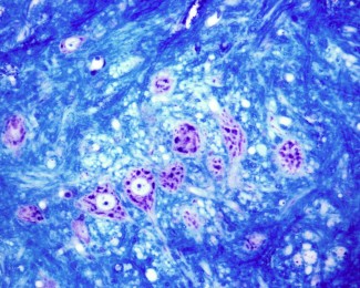
Spinal injuries are rarely anatomical ruptures with loss of continuity, but instead tend to be contusions. Although a lot of clinical research teams are studying these injuries, we still do not know how to repair the spinal cord.
In the case of tetraplegia or paraplegia, the aim is to enable the severed or compressed spinal cord to function again by restoring the continuity of the millions of axons that run through it.
A high number of research avenues have been explored in an attempt to improve the protection of the spinal cord; in other words, the progression and extension of the lesion after injury (secondary lesion of the spinal cord).
Research by Paris Brain Institute
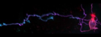
Claire Wyart’s team is developing fundamental approaches to spinal cord repair using zebrafish, due to their exceptional regeneration capacities. She is seeking to understand how the nerve networks of the spinal cord are recruited to carry out a series of complex locomotor actions.
For example, the spinal locomotor network (or rhythm-generating centre), of particular interest to the team, enables us to walk without thinking about it once we have decided to move. This ability to maintain movement comes from the network’s capacity to generate electrical oscillations. A range of data (physiological, pharmacological and anatomical) has been used to understand how these oscillations are generated in vitro (outside a living organism). However, these approaches do not reveal whether discharges from a given sub-group of neurons are necessary and sufficient to generate movement.
To compensate for this, the team is studying the function of specific spinal cells in vivo in larval zebrafish. Although this animal model may seem far removed from humans, it offers key advantages for research that in the long term aims to repair the spinal cord and restore normal locomotion in patients with disabilities. The zebrafish nervous system evolves very rapidly, which helps us understand these mechanisms quicker. What’s more, the larvae are transparent, making them particularly well suited to optogenetics, a cutting-edge technique that uses light to trigger targeted neurons from a distance.
This new approach makes it possible to activate and deactivate sub-groups of neurons and determine their role in the fish’s movement. This allows us to dynamically test the genetic role of an identified neuron type in the initiation and modulation of locomotor behaviour in an awake animal. The team aims to determine how sensitivity to movement contributes to locomotion, and it recently discovered that, in addition to neurons that contact cerebrospinal fluid (CSFns), there are mysterious sensory neurons in the centre of the spinal cord. Their activation modulates the movement of the larva. This observation opens up a new field of investigation into the sensory interface between the spinal cord and the cerebrospinal fluid.
Professor Hugues Pascal-Moussellard is an orthopaedic surgeon at Pitié-Salpêtrière Hospital, specialising in spinal column surgery. He is involved in research at Paris Brain Institute in collaboration with Dr Claire Wyart. Their research work aims to make progress in addressing the problem of myelin repair (the protective sheath that surrounds axons and enables nerve information to be transmitted) in patients with spinal cord injuries.
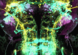
The Sensorimotor Integration Brain & Body Integration Lab team is studying neuromodulatory networks originating from the brain or spinal cord and their effects on locomotion and posture. The team is interested in reticulospinal neural circuits (RSNs)...
Read moreSupport Paris Brain Institute
Did you like this content and did it answer the questions you were asking? Don't hesitate to support Paris Brain Institute.