Scientists have access to several brain scanning methods to better understand the brain and how it works. Here we provide an overview of the techniques used at Paris Brain Institute.
Electrophysiology
Electrophysiology records the electrical signals (nerve impulses or action potential) emitted by neurons to communicate with each other. It is essential that we understand how the brain works under normal conditions, to understand and better treat impaired functions in nervous system disorders and also to preserve the brain in its normal state. The electrical signal transmitted by each neuron after activation is a key component of brain activity that is altered in neurological and psychiatric diseases, and is often the cause of the deficits observed.
Disruptions to action potentials are, for example, the ‘starting point’ for epilepsy. At Paris Brain Institute, there is specific research to investigate extreme brain conditions that exhibit continuous and abnormal electrical activities.
Neuroimaging
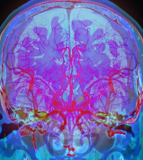
Because of advances in neuroimaging in recent decades, there has been a significant increase in knowledge about anatomy and how the nervous system works. Neuroimaging techniques can be grouped into two main categories:
- Neurophysiology techniques based on measuring the electrical or magnetic activity of brain cells, such as electroencephalography (EEG) and magnetoencephalography (MEG).
- Techniques that indirectly measure changes in brain activity through changes in brain perfusion or by injecting radioactive molecules, such as magnetic resonance imaging (MRI) and positron emission tomography (PET).
Neuroimaging research at Paris Brain Institute focuses on three main areas:
- Clinical research: studying major diseases of the nervous system and developing innovative treatments;
- Cognitive science research: understanding how the brain works and studying the neural bases for thought, behavior and aging;
- Signal and image processing research: developing new methods for acquiring and processing brain activity and imaging data.

Magnetoencephalography
Molecular and cellular techniques
Molecular and cellular techniques are used to understand the genetic, molecular and cellular bases of central nervous system development and function, and central nervous system disorders. These include:
- Genetic sequencing. This means reading the long DNA molecules that form chromosomes. This reading makes it possible to analyze the genome, detect any gene mutations and identify possible links between these mutations and the emergence of neurological diseases.
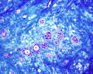
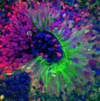
- Cellular investigations, by establishing easily manipulable cell cultures to create a simplified reproduction of the mechanisms of nervous system conditions. This work requires the activity of neuronal cells to be recorded, to identify any anomalies in the transmission of the electrical signal, and to manipulate ‘stem’ cells that become pluripotent to produce authentic nerve or glial cells. When examining function or impaired function of the brain as a whole, histology techniques on sections of tissue can evaluate the integrity of neuron and glial cell populations within different regions of the brain.The structures of the brain can also be visualized in 3D, on brain tissue made transparent through a technique known as CLARITY imaging.
- Cell imaging, to observe molecules, cells and tissue sections, cellular movements or even compartments within cells at microscopic scale (organelles, viruses, crystals, molecules).
AI-driven brain research
Paris Brain Institute is working on the development of new mathematical and computational approaches to studying the structure of the human brain and its functional networks. Transforming raw imaging data into formalized models such as geometric models of brain structure, statistical models of populations and connectivity graphs is now essential to defining new biomarkers of disease, studying the correlations between genetics and symptoms, and identifying the functional responses of the brain.