Thanks to 7T fMRI, researchers from Paris Brain Institute and NeuroSpin, the CEA's neuroimaging centre, are exploring the neural substrate of visual imagery at very high resolution for the first time. Their results, publiés[i] in Cortex, pave the way for a better understanding of this fascinating cognitive ability, which some of us entirely lack.
Visual imagery—the ability to mentally summon the image of a landscape, a person, or an object that is not directly observable—varies greatly in intensity from one individual to another. Some people can recall a detailed city map and walk through each street as if watching a movie. Thinking of a loved one, others may barely make out their silhouette and hair color.
Interestingly, about 4% of the population seems completely unable to visualize a scene on demand: this is known as aphantasia, a cognitive peculiarity known for over a century but only recently studied by scientists.
Preliminary studies suggest that aphantasia is present from birth and often affects multiple members of the same family. While it is not considered a disorder, it is frequently associated with a weaker-than-average autobiographical memory, difficulty recognizing faces, or even autism spectrum disorder. However, these associations remain uncertain and hard to explain.
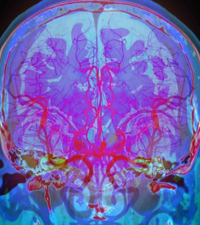
To understand what characterizes aphantasia at the brain level, we needed to study the neural mechanisms involved in visual imagery and perception. For this, we took advantage of 7-Tesla fMRI, which allows us to observe brain activity at extremely high resolution.
Most previous studies on aphantasia relied on highly subjective tests in which participants had to self-evaluate their visualization abilities. Under such conditions, it’s difficult to determine whether these tests actually assess mental imagery or instead reflect metacognition, that is, a person’s ability to describe their own mental processes.
In collaboration with Stanislas Dehaene's team at NeuroSpin, the CEA's brain imaging center, postdoctoral researcher Jianghao Liu, Paolo Bartolomeo, and their colleagues sought to examine the characteristics of aphantasic individuals more objectively.
We wanted to identify the precise neural circuits involved in mental imagery and visual perception. Most importantly, we aimed to understand how visual information is processed in the brain in the absence of visual stimuli.
Visualizing a scene with the “mind’s eye” is cognitively complex. It involves experiencing an object's visual properties without the object being present. This process not only recruits brain circuits linked to sensory experience but also draws on language and memory.
To break down this process, the researchers recruited 10 aphantasic subjects and 10 subjects with “typical” mental imagery. These individuals underwent ultra-high-field functional MRI while responding, from memory, to questions about the visual features of familiar objects, words, faces, and places.
The study found that attempting mental visualization activates fronto-parietal networks, which are important for attention, awareness, and working memory, as well as the left fusiform gyrus, located on the underside of the temporal lobe. It also activates areas of the ventral temporal cortex involved in letter recognition, face perception, and color processing.
Among aphantasic individuals—even if they report no experience of mental imagery—these same regions were also activated, but with reduced functional connectivity. In other words, these areas communicated less efficiently than in people with typical mental imagery.
These preliminary findings support a hypothesis already considered by researchers: the quality of visual experience, whether based on perception or imagination, depends on how well information is integrated between fronto-parietal networks and visual perception networks. The left prefrontal cortex might play a causal role in the awareness of these visual experiences.
“This might explain why aphantasic people still retain accurate visual knowledge of objects. For example, they clearly remember that spinach is a darker green than lettuce,” Liu notes.
Future studies will help determine whether aphantasia manifests in the same way for all affected individuals or whether there are subtypes linked to different causes.
In addition to highlighting the extreme variability in how we experience the world, research on aphantasia shows that mental imagery is not a prerequisite for reasoning, imagination, conceptualization, or creativity. Ultimately, these studies may illuminate the relationships between mental imagery, perception, memory, and neurodevelopment.
Sources
Financement
This study was funded by Dassault Systèmes.
Références
[i] Liu, J., et al. Visual mental imagery in typical imagers and in aphantasia: A millimeter-scale 7-T fMRI study. Cortex, April 2025. DOI: 10.1016/j.cortex.2025.01.013
[1] Liu, J., Bartolomeo. P. Aphantasia as a functional disconnection. Trends in Cognitive Science, June 2025. DOI: 10.1016/j.tics.2025.05.012

Consciousness, attention, visual perception and language are complex cognitive functions involving different brain areas and neural networks.
Read more