First described 60 years ago, chronic myoclonus following cerebral anoxia is now known as Lance-Adams syndrome. This is a severe disorder whose mechanisms were, until now, poorly understood. Geoffroy Vellieux, Vincent Navarro, and their colleagues at Paris Brain Institute now show that this condition originates in the motor cortex. Their findings are published in the journal Neurology.
Lance-Adams syndrome (LAS) is a severe disease that occurs after prolonged oxygen deprivation to the brain—known as cerebral anoxia—such as after a cardiac arrest. When patients regain consciousness after a stay in intensive care, they suffer from muscle jerks called myoclonus, which occur at rest or during movement. These symptoms interfere with daily activities and can cause frequent falls. Most importantly, they persist over time—resulting in long-term disability.
This syndrome is relatively rare, affecting only 0.5% of patients who present with myoclonus after cerebral anoxia. Even though we've known about this syndrome for a long time, the underlying pathological mechanisms remained a mystery
The Largest Cohort Ever Assembled
Understanding the pathophysiology of the disease is essential for developing effective therapeutic approaches. But in the case of Lance-Adams Syndrome, researchers faced a major challenge: the patients' muscle jerks are very brief (a few milliseconds), sometimes barely noticeable, making their observation and clinical interpretation difficult.
Moreover, although electroencephalography (EEG) had already been used by clinicians in the earliest studies on LAS in the 1960s, it does not allow precise identification of the origin of brain abnormalities.
To shed light on these uncertainties, researchers from the “Clinical and Experimental Epilepsy” team, co-led by Prof. Vincent Navarro (AP-HP, Sorbonne University), studied 18 patients treated at Pitié-Salpêtrière Hospital—a significant cohort for a rare disease and the largest ever assembled for this condition.
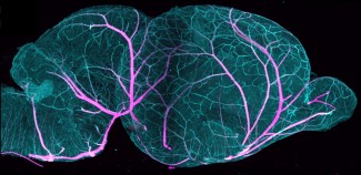
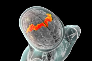
Thanks to extensive neurophysiological and neuroradiological examinations (including electromyography, electroencephalography, and positron emission tomography), the team showed that the patients’ myoclonus predominantly originates in the cerebral cortex, specifically in the motor cortex.
“These results confirm the initial intuition of Raymond Adams and James Lance, the neurologists who gave the disease its name in 1963,” notes Vincent Navarro.
New Therapeutic Avenues
Within the cohort, 11 patients also experienced epileptic seizures. Surprisingly, for 8 of them, myoclonus was significantly reduced following these seizures. “Based on this observation, we proposed electroconvulsive therapy to one patient—which involves delivering an electrical stimulus through the brain—to reproduce the seizures in a controlled way. The treatment significantly alleviated her symptoms, which had been resistant to medication,” the researcher adds.
This finding paves the way for new strategies to modulate abnormal cortical activity.
Finally, the researchers plan to carry out new studies to pinpoint exactly which neuron populations and networks, within the motor cortex, trigger the cascade of signals that causes myoclonus—with the goal of developing new treatments, drug-based or otherwise, to improve patients' quality of life.
Funding
This study was funded by the "Investments for the Future" program and the AP-HP Foundation.
Sources
Vellieux, G., et al. Multimodal Assessment of the Origin of Myoclonus in Lance-Adams Syndrome. Neurology, Décembre 2024. DOI: 10.1212/WNL.0000000000209994.
Vellieux G., et al. Effectiveness of electroconvulsive therapy in Lance-Adams syndrome. Brain Stimulation. Mars 2023. DOI: 10.1016/j.brs.2023.03.004.

The team "Clinical and experimental epilepsy" explores how the brain becomes epileptic (epileptogenesis), how it produces seizures (ictogenesis) and the relationships between neurophysiological activities and clinical semiology. The team is also...
Read more