
Grasping an object, walking, driving, writing: all the voluntary movements required for these actions are controlled by the brain.
How the brain controls voluntary movements
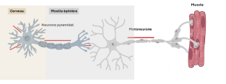
Voluntary motor skills are controlled by what’s known as the pyramidal pathway. This pathway is made up of pyramidal or corticospinal neurons (from the motor cortex to the spinal cord) and motor neurons (from the spinal cord to the muscles).
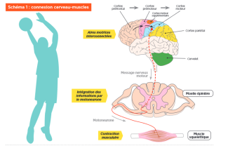
Voluntary movements happen thanks to sequential or simultaneous activation of different regions of the brain (diagram 1):
- the prefrontal cortex and parietal cortex assimilate information needed to plan the movement according to the surroundings;
- the premotor cortex and additional motor cortex organize the sequence and coordination of complex movements;
- the motor cortex sends the command to the muscles to perform the gesture;
- the basal ganglia and
These brain areas are interconnected by networks of neurons that allow them to communicate, and this dialogue evolves according to the individual movement performed. There is a motor cortex in both hemispheres of the brain, each controlling the opposite part of the body: the motor cortex of the right hemisphere controls movements on the left side of the body, and vice versa.
Through communication and synchronization between the different regions according to their importance in the planned movement, a gesture can be adapted to a person’s surroundings.
There are differences between physical activities in reproducible environments like running, and in more unstable environments like team sports, where the position and movements of each player need to be assimilated.
The networks of neurons that allow the different regions of the brain to communicate are plastic (neuroplasticity). Learning and practicing a physical activity, in other words particular motor sequences, leads to morphological and functional changes in the brain.
The cerebellum is a part of the brain located at the back of the brain stem. Though small in size, it has all the brain’s characteristics with a cortex, deep nuclei and white matter. It also contains around half of the brain’s neurons. The cerebellum plays a key role in motor control, and particularly in using sensorimotor information to adapt and coordinate each movement. It is also important for balance. We also now know that the cerebellum is involved in several cognitive functions such as attention, language and emotions.
Also known as basal nuclei, the basal ganglia is a system of interconnected gray matter nuclei located deep within the brain. These structures extract information from the cortex to be selected, reinforced, learned and automated.
Motor skills at Paris Brain Institute
At Paris Brain Institute, the goal of the FORT[É] collaborative project is to identify and describe the brain circuits involved in the motor learning of complex movements. The researchers are seeking to establish a causal link between how fine motor skills are learned and the connectivity between the cerebellum and motor areas, through electroencephalogram (EEG) recordings and functional MRI. The results of this project will help to optimize physical therapy programs for stroke and brain injury patients, by modulating the neural circuits involved in movement through transcranial magnetic stimulation (TMS). Methodological and theoretical advances resulting from this research could also be applied to patients suffering from dystonia, Parkinson’s disease and epilepsy following epileptogenic foci surgery. To fully understand the dynamics and outcomes of motor recovery, it is essential to consider the physiological processes of motor skills learning when planning personalized training programs for patients with neurological disorders.
La motricité est la capacité du cerveau à organiser et exécuter les mouvements volontaires du corps.
La motricité dépend de plusieurs régions cérébrales : le cortex moteur, le cortex prémoteur, le cervelet et les ganglions de la base.
Le cortex moteur envoie les commandes nécessaires à l’exécution des mouvements vers les muscles.
Le cervelet permet de coordonner les mouvements et d’ajuster leur précision en fonction des informations sensorielles.
Les ganglions de la base participent à la sélection des mouvements et à leur automatisation, en facilitant les gestes adaptés et en inhibant les mouvements parasites.
Grâce à la plasticité cérébrale, les réseaux neuronaux impliqués dans la motricité se modifient avec l’apprentissage et l’expérience.
Au cours des dernières années, de nombreuses études neuroscientifiques ont porté sur la relation entre le sport et les capacités cognitives dans différentes populations, des plus jeunes aux plus âgés.
Cliquez ici