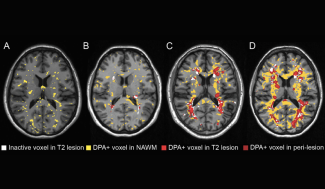
Imaging technique that measures the metabolic activity of a tissue using radioactive compounds.
Positron emission tomography (PET) is based on the intravenous injection of a substance (the “tracer”) labeled with a radioactive atom, fluorine 18 or carbon 11, which, on attaching itself to the target cells, emits specific particles known as positrons. These particles then collide with electrons, producing photons (light particles). The tracer is chosen to bind to a specific organ or tissue, enabling us to reconstruct an image of the organ under study - in this case, in MS, the brain. The radioactive substances used in a PET scan are harmless to humans, and the very low levels of radioactivity disappear completely within 1 day.
This technique makes it possible to visualize directly in vivo and in real time, the kinetics and distribution of injected radiotracers, and therefore of the molecules to which they bind. It is often combined with an MRI performed on the same machine to obtain more precise images of the organs studied.
- Brain Imaging :
- Means all medical imaging techniques that can be used to observe the brain (MRI, Scanner, EEG, MEG-EEG, etc.).
- Magnetoencephalography (MEG) :
- Technique for measuring magnetic fields induced by the activity of neurons in the brain or spinal cord.

