
Lower part of the central nervous system located in the vertebral column.
The spinal cord, with the brain, forms the central nervous system. It is a cord located in the spinal canal that extends from the brain stem to the second lumbar vertebra. Spinal cord injuries can lead to numerous conditions requiring appropriate treatment.
The main function of the spinal cord is to transmit nerve signals between the brain and the rest of the body. It enables the transfer of motor information to the muscles and sensory information to the cortex, and includes a coordination center for certain reflexes that are independent of brain control.
What is the Difference Between the Spinal Cord and Bone Marrow?
The spinal cord should not be confused with bone marrow or hematopoietic marrow. The latter is located inside the bones and its role is to transform stem cells into blood cells (white blood cells, red blood cells, and platelets). Changes in the bone marrow can lead to serious blood disorders.
What is the Structure of Spinal Cord?
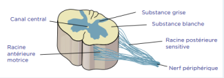
The spinal cord is protected by the vertebral column (spine), which consists of 7 cervical vertebrae (C1-C7), 12 thoracic vertebrae (T1-T12), 5 lumbar vertebrae (L1-L5), and 5 sacral vertebrae (S1-S5).
Two roots emerge from each intervertebral space (there are 31 in total), one motor and one sensory, corresponding to a specific region of the body.
Nerves are composed of bundles of axons (roots), which are extensions of neurons, and each bundle transmits a nerve signal that travels up to the brain (ascending) or comes from the brain (descending). The spinal cord generates 8 cervical spinal nerves (C1 to C8), 12 thoracic spinal nerves (T1 to T12), 5 lumbar spinal nerves (L1 to L5), 5 sacral spinal nerves (S1 to S5), and 1 coccygeal spinal nerve (C1).
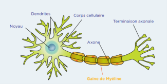
A neuron consists of a nucleus, dendrites, a cell body, one or more axons covered with myelin, and an axon terminal.
The Motor Roots
The motor roots are located at the front of the spinal cord in the anterior horn. Information, or nerve impulses, travels down from the brain to the rest of the body. These roots enable voluntary movement. At each intervertebral space, motor nerve fibers (nerves) emerge. These nerves are specifically connected to parts of the body but can control the contraction of several muscles.
There are five plexuses, which are fusions of several nerves:
- The cervical plexus, formed by the fusion of the cervical nerves from C1 to C5, connected to the muscles of the neck and shoulders
- The brachial plexus, which groups together the cervical and thoracic nerves from C5 to T1 and is connected to the muscles of the arms and upper back
- The lumbar plexus formed by the lumbar nerves from L1 to L4, which controls the muscles of the abdomen and legs
- The sacral plexus, composed of the lumbar and sacral nerves from L4 to S4, connected to the muscles of the legs
- The coccygeal plexus, which merges the sacral nerves S4 that control the genitals and sphincters.
The Sensory Roots
The sensory roots are located at the rear of the spinal cord in the posterior horn.
Information, or nerve impulses, travels up from the body towards the brain. These roots enable the brain to receive information about touch, body and limb position, pain, vibrations, and temperature from the body. Sensory nerve fibers (nerves) emerge from each intervertebral space, originating specifically from different areas of the skin called dermatomes. A dermatome is an area of skin with sensory nerves that all originate from a single root. For example, sensory information from the back of the thigh is transmitted to the brain through sensory nerves entering the spinal cord through the foramen of the second sacral vertebra (S2).
What are Spinal Cord Injuries and Pathologies?
Spinal cord injuries, whether traumatic or chronic, cause symptoms specific to the affected area. These injuries can cause loss of sensation, changes in reflexes, motor deficits, and even paralysis (paraplegia or quadriplegia).
Damage can be caused by a pinched nerve root at the exit point from the spinal column, a tumor, an infection, a traumatic injury following a fall or accident, or a chronic condition such as multiple sclerosis or ALS.
The diagnosis is made through clinical examination of the impaired functions, which makes it possible to identify the damaged area of the spinal cord, and through MRI.
Treatment for spinal cord injuries is tailored to their cause, involving surgery for bone-related causes and drug therapy for chronic or infectious diseases.
At Paris Brain Institute
The Spinal Sensory Signaling team is looking into the role of the spinal cord in locomotion
The art of wandering in vertebrates: new mapping of neurons involved in locomotion
Amyotrophic lateral sclerosis (ALS or Motor Neuron disease) or spastic paraplegia are conditions caused by degeneration of the corticospinal neurons that control voluntary movement.
The Causes of ALS and Mechanisms of Motor Neuron Degeneration team aims to better understand and halt the death of spinal motor neurons. Therapeutic approach to ALS or Motor Neuron disease
The Basic to Translational Neurogenetics team is seeking to identify the causes and new treatments for spastic paraplegia
In multiple sclerosis, certain demyelination lesions are located in the spinal cord. Two research teams are working to repair these lesions:
- The Repair in Multiple Sclerosis: from Biology to Clinical Translation team.
- The Myelin Plasticity and Regeneration team.
Early cortical remyelination has a neuroprotective effect in multiple sclerosis