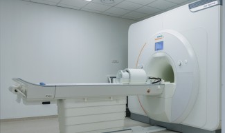
This week, Paris Brain Institute inaugurated an exceptional piece of equipment: the latest generation 7-Tesla MAGNETOM Terra.X MRI scanner from Siemens Healthineers. This acquisition, made possible thanks to the support of RICHARD MILLE and the commitment of the Paris Region, is part of an ambitious investment plan for the Institute's neuroimaging facility. With this 7T MRI scanner, the Institute is taking a technological leap forward for the benefit of the neuroscience community and, ultimately, for patients.
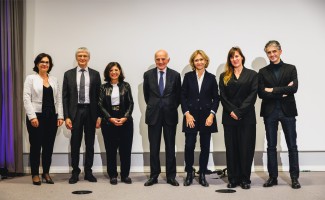
Paris Brain Institute's 7T MRI scanner was inaugurated on 25th November in the presence of Valérie Pécresse, President of the Paris Region, Timothée Malachard, Marketing Director of RICHARD MILLE, Amanda Mille, Brand and Partnership Director of RICHARD MILLE, and members of the Institute's Board of Directors. This is the fifth ultra-high-field MRI scanner in France and the only one located in the heart of a university hospital in the Paris region.
“The decision to equip Paris Brain Institute with a 7T MRI scanner dates back to the creation of the Institute in 2010,” Alexis Brice, Managing Director of the Institute, explains. “This is an emblematic investment for highly ambitious research. This machine will reinforce a vast network of similar equipment throughout, the strength of which is closely linked to the advancement of knowledge in neuroscience.”
Richard Mille's long-standing and loyal support of Paris Brain Institute’s teams—since its creation—testifies to our commitment to supporting ambitious scientific projects. We believe that they can shape the future of biomedical research. As we celebrate the inauguration of this 7T MRI scanner, we picture how this technological milestone will positively impact the neuroscience community.
Paris Brain Institute is the first user in France to benefit from the latest advances in MAGNETOM Terra.X technology, which the manufacturer is constantly improving. Thanks to dynamic signal homogenization techniques, powerful gradients and multi-core imaging capabilities, the CE-marked system will provide images of unrivalled precision at 7T—opening up new applications such as very high-resolution imaging, sodium imaging in brain tumors and precise tracking of nerve fiber bundles.
Exploring the brain at the sub-millimeter level
7T MRI technology represents a qualitative and quantitative leap forward in spatial resolution and morphological, functional and metabolic imaging.
“Thanks to 7T MRI, it is possible to thoroughly observe structures a few hundred microns in size, much smaller than those previously visible with 3T MRI,” Brian Lau, Scientific Director, adds. “Cortical anatomy has traditionally been studied using microscopic imaging of post-mortem tissue. However, using a non-invasive technique such as the 7T MRI scanner makes it possible to map the brain in an extremely precise, micro-anatomical way. Ultimately, we could identify new therapeutic targets and expand the use of deep brain stimulation.”
“7T MRI is more precise than the 1.5 or 3 Tesla MRI scanners commonly used in hospitals,” Jean-Christophe Corvol, Medical Director, says. “It allows us to characterize small structures, such as the brainstem, which connects the brain to the spinal cord, and to study its anatomical sub-sections. The 7T MRI scanner will also make it easier to subdivide the cohorts of patients followed at the Institute as part of studies on neurodegenerative and neurogenetic diseases.”
A hub of neuroimaging expertise in the Paris region
The acquisition of this scanner was made possible thanks to the exceptional support of RICHARD MILLE. It is part of a major project to create a new “SESAME” track, a program to structure strategic industries in the Paris Region. It is based on the scientific and technological capacities of universities and research organizations and is funded by the Paris Region and the French government as part of the “France 2030” Plan.
The Neuro@7T project led by Paris Brain Institute is the winner of the third “SESAME filières France 2030” call for proposals. It aims to develop a new hub of expertise in ultra-high-field MRI neuroimaging in the Paris Region, identify innovative diagnostic, prognostic and follow-up imaging markers for therapeutic trials, and ultimately benefit patients suffering from neurological and psychiatric disorders.
By contributing to the funding of this equipment at Paris Brain Institute through the SESAME program, we have chosen to enable neuroscience research in the Paris Region to remain internationally competitive and innovative. I want to strengthen the Paris Region ecosystem with breakthrough technologies and make it Europe's Eldorado for innovation. This inauguration is the fruit of this policy, which we have enshrined in our 2025 budget.
Neuro@7T will also offer a healthcare track to deploy new digital technologies and methods to support diagnosis and care for academic and industrial neuroscience partners.
The project builds on the technological expertise of the CEA-Neurospin research Center—a world expert in ultra-high-field neuroimaging—and the leadership of Paris Brain Institute in clinical and translational neuroscience. These two organizations will structure a network of leading research institutes in basic and translational research in the Paris Region.
A wide range of research possibilities
The devices currently available on Paris Brain Institute's imaging platform—such as the latest-generation 3T MRI scanner installed in March 2024, PET-MRI, MEG-EEG and now the 7T MRI scanner—are designed to complement each other. It opens up new research perspectives for the development of precision medicine by individualizing care pathways and therapeutic procedures.
Thanks to its location at the heart of the Pitié-Salpêtrière Hospital and the medical, scientific and technological expertise of its teams, Paris Brain Institute aims to accelerate the discovery of new prognostic and predictive markers for brain and spinal cord diseases.
Ultra-high-field MRI opens up promising avenues for clinical imaging in multiple sclerosis, brain tumors, epilepsy, neurodegenerative and neurovascular diseases. However, basic research, the cornerstone of Paris Brain Institute's scientific strategy, will not be left behind.
Studying the specialization of certain brain regions for reading, investigating the metabolic processes associated with mental fatigue, exploring the basal ganglia in minute details... these new research projects would not have been possible without the 7T MRI, and will help us understand how the brain works.
Finally, in collaboration with the Montreal Neurological Institute-Hospital in Canada, the Institute's researchers will use the new equipment to build up a massive database of healthy subjects based on a cohort of 600 people aged between 8 and 80. This database will be accessible to researchers worldwide, with an open science approach.
