Depression results from a malfunction in the transmission of information from one neuron to another in the brain. Between each pair of neurons, there is a gap called a synapse, in which the neuron transmitting information releases molecules called neurotransmitters. These molecules are then captured by the neuron receiving the information. The uptake of neurotransmitters by a neuron triggers the generation of an electrical current, known as a nerve impulse or action potential, which travels along the axon, the extension of the neuron, to the next synapse, where it triggers the release of neurotransmitters.
Biological Mechanisms of Depression
A disruption in the production and uptake of three major neurotransmitters is responsible for the development of major depressive episodes.
- Serotonin, which regulates sleep, appetite, and mood
- Dopamine, which regulates mood and motivation
- Noradrenaline, which regulates attention and sleep
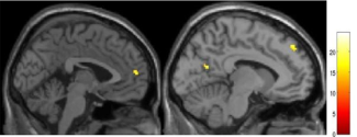
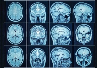
These neurotransmitters are present in many areas of the brain. Functional brain imaging (functional MRI) can identify the areas of the brain most involved in depression.
Depression is a mood disorder that significantly impacts brain network dynamics involved in emotional regulation, cognitive control, and self-referential processing.
At Paris Brain Institute
The French National Research Agency (ANR) project “SENSO” coordinated by Prof. Philippe Fossati has made it possible to define brain imaging markers that facilitate the diagnosis of depression. Functional MRI helped identify a malfunction in a network connecting different regions of the brain. Under normal conditions, this neuronal network is responsible for determining which external stimuli are worthy of attention.
Among other things, this project has highlighted the role of the medial prefrontal cortex, which plays a role in sadness and self-deprecation, the left dorsolateral prefrontal cortex, which is involved in working memory, and the precuneus, which is associated with rumination. It also highlighted the role of the ventral cingulate cortex in sensitivity to social exclusion signals, a key factor in triggering a major depressive episode.