A giant billboard flashes on the side of a country road. Why does it catch our attention more easily than other details in the landscape? At Paris Brain Institute, Tal Seidel Malkinson, Jacobo Sitt, Paolo Bartolomeo, and their colleagues show that exogenous attention—the ability to be involuntarily attracted to a specific element in our environment—is built up in the cortex gradually, from the back to the front of the brain, within three fronto-parietal networks. These neural networks allow us to explore space efficiently by disregarding familiar objects and favouring new or unexpected visual stimuli. The researchers' findings are published in the journal Nature Communications.
In a world inundated with a constant stream of new information—notifications, ads, emails, news—we often struggle to prevent our attention from being constantly hijacked by external events. But is it truly within our power to filter and select our perceptions? And why do we find ourselves so easily distracted?
“Exogenous attention, the cognitive process that allows a salient visual stimulus to impose itself on us, is automatic. When a colleague walks past our desk, our attention is diverted from our computer screen despite ourselves,” explainsTal Seidel Malkinson (University of Lorraine), a former postdoc at Paris Brain Institute and now a professor and researcher in neuroscience. “This phenomenon is all too familiar to anyone who tries to stay focused. Yet, the brain mechanisms behind it are still poorly understood.”
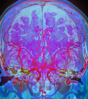
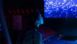
Functional MRI or electroencephalogram (EEG) are usually used to study the neural substrate for exogenous attention, but these techniques are limited by poor temporal or spatial resolution. “To track how the brain constructs spatial attention—which involves large and fast neural networks—we needed to record the electrical activity of neurons throughout the cortex and in great detail,” the researcher adds.
As close to neurons as possible
To this end, Malkinson and her colleagues recruited 28 patients who received depth electrodes as part of a pre-surgical assessment for drug-resistant epilepsy. Placed individually for each patient, the electrodes covered about 1,400 contact zones deep in the brain, giving the researchers a detailed view of neural activity during attention tests.
Participants had to look at two boxes separated by a small cross designed to focus their attention. Occasionally, a target appeared in the box on the left or right; the subject had to press a button to signal that they had seen it. Finally, the targets were preceded by peripheral visual cues capable of capturing the subject's attention and announcing where the target was about to appear (valid cue) or the opposite area (invalid cue).
This protocol is useful for measuring under which conditions the attention is captured by one event or redirected towards another. It also makes it possible to measure the subjects' reaction time and understand which visual stimuli the brain is likely to group—to process them either as a single event or as successive events.
To interpret the highly complex data from the intracerebral recordings, Tal Seidel Malkinson and Jacobo Sitt (Inserm) have developed an unsupervised learning method: an algorithm groups the electrodes that have a similar activity over time to reveal the global dynamics of the studied areas. This approach enables researchers to observe brain activity while reducing the influence of their theoretical preconceptions.
The gradient of attention in action
Their results identified three cortical networks that were successively activated from the back to the front of the brain when the participants' attention was captured by visual stimuli, as if attention were gradually developing in the cortex until the subject's final reaction—in this case, pressing a button.
“There is a continuum of activity in the cortex. In networks identified in the parieto-occipital regions, brain activity first processes visual information. Then, in the frontal regions, it reflects the behavioural response”, Malkinson explains. “We have shown that attention emerges like a bridge between these two poles. In a way, attention connects perception to action!”
“This is the first time the dynamics of exogenous attention networks appear so clearly, as well as their place in the organisation of the cortex”, Bartolomeo says. “Moreover, this work allowed us to observe the neural correlate of inhibition of return—an attentional phenomenon in which a longer reaction time is observed when someone is exposed to visual stimuli in a region of space they already explored, compared with a region of space that is still unknown.”
Inhibition of return is a filter that allows us to ignore familiar visual information automatically. For example, if we are trying to spot a squirrel in a tree, the branches we have examined will no longer be the focus of attention, even if they are waving in the wind.
Inhibition of return probably promotes efficient exploration. It is frequently defective in some patients who have suffered a stroke. Increasing our knowledge of the mechanisms of attention could, in the long run, contribute to treating them better.
Funding
This study was funded by the Israel Science Foundation, the Marie Sklodowska Fellowship and the French National Research Agency (ANR).
Sources
Seidel Malkinson, T. et al. Intracortical recordings reveal Vision-to-Action cortical gradients driving exogenous attention. Nature communications, (2024).
DOI : 10.1038/s41467-024-46013-4.