Or 34,00 After 66% tax deduction

I make a monthly donation I make an IFI donation
Research, science & health

First demonstration of the neuroprotective effect of remyelination in multiple sclerosis patients

Published on: 24/02/2022 Reading time: 1 min
First demonstration of the neuroprotective effect of remyelination in multiple sclerosis patients

In an article recently published in the European Journal of Neurology, Vito Ricigliano (AP-HP), Benedetta Bodini (AP-HP/Sorbonne University) and their collaborators at the Paris Brain Institute, demonstrate the protective effect of myelin repair on the tissues surrounding lesions in patients with multiple sclerosis. This discovery underlines the potential of new therapeutic strategies and provides new elements to evaluate the efficacy of remyelinating drugs currently being tested.

How to prevent or reduce neuronal degeneration, which is the cause of clinical disability in multiple sclerosis (MS)? At present, doctors have treatments available to control the inflammatory component of the disease but are powerless to deal with the degenerative component.

The study of experimental models has shown that myelin repair can protect the integrity of neurons and prevent neurodegeneration, which spreads from the demyelinating lesion all along the nerves that are not yet demyelinated but will degenerate directly

Benedetta Bodini Neurologist and last author of the article

In fact, in multiple sclerosis, the damage to the neurons is not only at the level of the myelin lesions visible on MRI, but extends to the regions surrounding them, the peri-lesional tissue. Researchers and clinicians at the Paris Brain Institute wanted to study whether spontaneous recovery of myelin - or remyelination - in the lesions could protect patients from microstructural damage to the surrounding tissue.

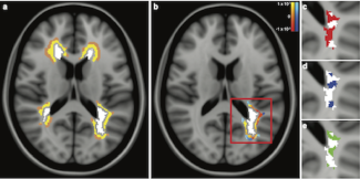
Lesions, perilesions and indices of myelin content change. a. Examples of lesional (white), proximal perilesional (yellow) and distal perilesional (brown) regions in the MRI of a patient with MS. b. White matter lesion (white) and its corresponding perilesions are represented with color maps, based on the delta of the mean diffusivity in each proximal and distal perilesional voxel from the first to the second time-point. c-e. Detail of the same white matter lesion of Fig. 1b, showing demyelinated voxels at

To do this, they monitored the amount of myelin in the lesions over time using PET-MRI and combined this with an assessment of the microstructural damage to the peri-lesional tissues using diffusion MRI, in 20 patients with multiple sclerosis. Their analyses were conducted at the single lesion level, i.e. over 500 lesions studied.

We show for the first time in vivo in MS patients that remyelination protects not only the lesion but also the surrounding tissue. This result highlights the importance of coupling existing anti-inflammatory therapies with a remyelination strategy to protect all tissues, even those that appear normal

Vito Ricigliano Neurologist and first author of the study

There is great heterogeneity among patients in their ability to remyelinate. In this study, the scientists also show that in the same patient, some lesions can repair themselves very well and others much less so, and that this difference is reflected in the damage to the peri-lesional tissue.


Clinical trials of remyelinating therapies are underway, notably in the team of Bruno Stankoff and Catherine Lubetzki at the Paris Brain Institute. Thanks to the use of PET-MRI, researchers will be able to study the effectiveness of the treatments, not only on the clinical signs, but also at the cellular level with the repair of myelin and the reduction of the microstructural damage to the surrounding tissues.

Sources

Spontaneous remyelination in lesions protects the integrity of surrounding tissues over time in multiple sclerosis. Ricigliano VAG, Tonietto M, Hamzaoui M, Poirion É, Lazzarotto A, Bottlaender M, Gervais P, Maillart E, Stankoff B, Bodini B. Eur J Neurol. 2022 Feb 12.

Our news on the subject

À la recherche de marqueurs d’imagerie dans la démence frontotemporale
Searching for Imaging Markers in Frontotemporal Dementia
Could exploring the relationships between different brain networks help us understand frontotemporal dementia (FTD)? This neurodegenerative disease, which progresses at varying rates, is often diagnosed late—when clinical signs are already severe. At...
01.07.2025 Research, science & health
Monocyte – un globule blanc qui se différencie en macrophage. Crédit : Université d’Edinbourg.
Discovery of a Macrophage Anomaly in Multiple Sclerosis
Certain patients with multiple sclerosis (MS) can partially regenerate myelin—the protective sheath that surrounds nerve fibers—which is damaged during the evolution of the disease. In studying how immune cells influence this remyelination...
12.19.2024 Research, science & health
Interneurones. Crédit : UCLA Broad Stem Cell Research Center.
Stimulating specific neurons in the striatum stops compulsive behaviour
What if we could resist compulsions? These irrational behaviours, particularly common in obsessive-compulsive disorder (OCD), are hard to suppress. At Paris Brain Institute, Éric Burguière's team shows that we can anticipate them and block them...
09.10.2024 Research, science & health
Les nerfs moteurs présents dans la moelle épinière se projettent vers la périphérie, où ils entrent en contact avec les muscles, formant des connexions appelées jonctions neuromusculaires. Crédit : James N. Sleigh.
Ultrasound show unexpected effects on motor neuron disease
Over the past fifteen years, neurosurgeons have been perfecting a fascinating technique: using ultrasound to temporarily open the blood-brain barrier to facilitate the action of therapeutic molecules in the central nervous system. At Paris Brain...
09.05.2024 Research, science & health
Un neurone
Rett syndrome: a new gene therapy on the way
Gene therapy could be our best chance of treating Rett syndrome, a neurological disorder that causes severe intellectual and motor impairments. At Paris Brain Institute, Françoise Piguet and her colleagues have looked closely at brain cholesterol...
07.16.2024 Research, science & health
Lésions d’un patient à l’inclusion dans le protocole (M0) disparues après 2 ans de traitement à la Leriglitazone (M24)
The dual effect of leriglitazone in X-linked Adrenoleukodystrophy (X-ALD)
In 2023, the team led by Professor Fanny Mochel (AP-HP, Sorbonne University), a Paris brain Institute researcher, showed that daily dose of leriglitazone slow down the progression of myelopathy in patients with X-linked adrenoleukodystrophy, and...
06.28.2024 Research, science & health
See all our news