
Or 34,00 After 66% tax deduction

I make a monthly donation I make an IFI donation
Research, science & health

New early marker in Parkinson’s disease

Published on: 29/01/2014 Reading time: 1 min
Le complexe coeruleus/subcoeruleus est visible dans cette nouvelle sequence IRM. Cela nous a permis d’étudier la relation entre cette petite structure (en blanc dans l’image) et le tonus musculaire pendant le sommeil paradoxal.
Retour à la recherche

A paper published in the prestigious journal Brain by a team of the Institut du Cerveau - ICM institute link rapid eye movement sleep disorders and Parkinson’s disease. These results open the possibilities to early diagnose development of the Parkinson’s disease.

Parkinson disease is a progressive degenerative neurological disorder affecting 150 000 persons in France. It is characterized by the death of a neuronal population producing dopamine, a neurotransmitter, chemical messenger allowing transmission of informations between neurons. The diminution of the concentration of dopamine is the source of the development of the disease. Indeed, symptoms arise when 60-80% of these specific neurons disappear.

These last few years, researchers studied non-dopaminergic symptoms and more notably rapid eye movement sleep behavior disorder affecting 30 to 60% of patients. Rapid eye movement sleep is traditionally characterized by dreams and is associated with a strong neuronal activity, ocular movements and diminution of muscle tone. Some patients with Parkinson disease have rapid eye movement sleep behavior disorders with increased muscular tone which could lead to nocturnal violence. Rapid eye movement sleep behavior disorders could arise in any healthy subject whithout neurological disease. However, these persons present a higher risk to develop Parkinson disease or demencia. Daniel GarcÍa-Lorenzo in the team of Marie Vidailhet et Stéphane Lehéricy just highlighted the role of a part of the brain named the coeruleus/subcoeruleus complex in the blocking of muscle tone during eye rapid movement sleep.

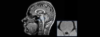
The team studied the integrity of this complex in patients with Parkinson’s disease using imaging approaches. They showed that patients with Parkinson’s disease have a reduced neuronal activity in the coeruleus/subcoeruleus complex associated with rapid eye movement sleep behavior disorder. Imaging datas combined with careful clinical evaluation as well as nocturnal video monitoring may be used as an early marker of non-dopaminergic Parkison’s disease pathology to predict the occurrence of Parkinson’s disease.

Le complexe coeruleus/subcoeruleus est visible dans cette nouvelle sequence IRM. Cela nous a permis d’étudier la relation entre cette petite structure (en blanc dans l’image) et le tonus musculaire pendant le sommeil paradoxal.
The coeruleus/subcoeruleus complex is visible in this new MRI sequence. This allowed us to study the relation between these small structures (in white on the image) and the muscle tone during rapid eye movement sleep.

Sources

Garcia-Lorenzo D, Longo-Dos Santos C, Ewenczyk C, Leu-Semenescu S, Gallea C, Quattrocchi G, Lobo PP, Poupon C, Benali H, Arnulf I, et al.: The coeruleus/subcoeruleus complex in rapid eye movement sleep behaviour disorders in Parkinson’s disease. Brain 2013, 136:2120-2129.

Our news on the subject

motoneurones
Parkinson's disease, multiple sclerosis, ALS: behind the rising case numbers, distinct dynamics
The number of patients living with neurodegenerative diseases that affect movement is rising steadily across Europe. Yet a large-scale study from the Paris Brain Institute and the Karolinska Institutet in Stockholm shows that this is not an emerging...
06.08.2026 Research, science & health
Microtubules in vitro. Crédit : Paul Appleton, Université de Dundee.
When physics sheds new light on brain diseases
For decades, cell biology has built its approach around the study of genes and molecules, pushing the physical forces that sculpt our tissues into the background. Today, an international consortium, coordinated by Fanny Mochel at the Paris Brain...
05.26.2026 Research, science & health
Rêver éveillé : les états oniriques ne sont pas réservés au sommeil
Dreaming while awake: dream-like states are not confined to sleep
We tend to take for granted that the thoughts associated with sleep have a particular quality: we often describe them as elusive, abstract, or marked by a certain strangeness. Yet a study conducted by researchers from the DreamTeam at the Paris Brain...
04.29.2026 Research, science & health
Représentation artistique des neurones. Crédit : Odra Noël.
How the architecture of the prefrontal cortex shapes our creativity
The cognitive and neural mechanisms underlying creative thinking are still poorly understood. A new study from the FrontLab team at the Paris Brain Institute explores this question from an original angle by examining creativity where it sometimes...
04.22.2026 Research, science & health
Des mini-cerveaux en laboratoire pour comprendre l'épilepsie de l'enfant
Lab-grown mini-brains shed light on childhood epilepsy
Why does the same genetic mutation cause a severe brain malformation in some patients but not in others? Researchers from the MOSAIC team at the Paris Brain Institute have developed mosaic human cortical organoids carrying mutations in the DEPDC5...
04.16.2026 Research, science & health
Comment les vaisseaux sanguins cérébraux se construisent après la naissance
How Brain Blood Vessels Develop After Birth
Researchers from the Paris Brain Institute and Sainte-Justine University Hospital in Montreal have, for the first time, revealed the key stages of vascular development in the brain, from birth through adulthood. Using a 3D digital atlas called...
04.15.2026 Research, science & health
See all our news