A collaboration between Australian (University of New South Wales and Macquarie University) and French (Paris Brain Institute) researchers has found that variations in the diameter of our pupil reveal the sensory intensity of our mental imagination, such as the brightness of an imagined scene. These results, published in ELife, also provide the first physiological validation of aphantasia, the absence of mental visualisation.
Closing your eyes and visualising a landscape, a picture, a face in your head... this faculty, visual or mental imagery, may seem obvious to many of us. However, it is not shared by everyone. It is estimated that around 3% of people are aphantasic, i.e. they do not possess this mental visualisation ability.
Today, visual imagery is often determined subjectively by asking a participant directly to describe their ability to visualise objects mentally. We lacked an objective tool to access an individual's mental imagery capacity.
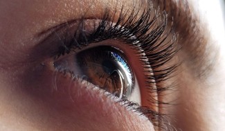
When we see an object with our eyes, our pupils adapt to it. Their diameter varies according to the brightness outside, but studies have shown that it also adapts to the interpretation of an image. Thus, the pupil’s diameter is smaller for a black-and-white image depicting a sunny beach than for a moon-lit landscape, even when the images were manipulated to have the same luminance: the eye uses the context to approximate the expected level of luminance.
The pupillary reflex is an adaption that optimises the amount of light hitting the retina. And while it was already known that imagined objects can evoke so-called ‘endogenous’ changes in pupil size, we were surprised to see more dramatic changes in those reporting more vivid imagery. This really is the first biological, objective test for imagery vividness.
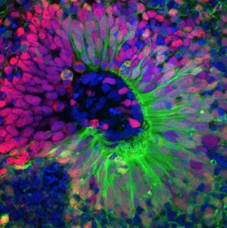
To explore this link between pupillary response and mental imagery, a group of Australian and French researchers set up two series of experiments. In the first, they presented participants with no visual imagery disorders with a series of geometric shapes varying in complexity and luminance, i.e. light intensity. The subjects were then asked to mentally visualise these shapes for 5 seconds. At the same time, their pupils were recorded using two cameras mounted on glasses. In a second phase, the researchers carried out the same study with aphantasic patients, who report no or limited mental imagery capacity.
The results show that the pupil diameter of the control subjects reacts both to the cognitive effort generated by the mental imagery and to the luminance of the visualised object. Furthermore, the pupil response to luminance was predictive of the subjective quality of the mental image reported by the subjects, i.e. how accurate they considered the image to be in their thoughts. In contrast, aphantasic subjects showed a smaller restriction of pupil diameter for more complex shapes but not for luminance.
Our pupils are known to get larger when we are doing a more difficult task. Imagining four objects simultaneously is more difficult than imagining just one. The pupils of those with aphantasia dilated when they imagined four shapes compared to one, but did not change based on the whether the shapes were bright or dark. This indicated that the participants with aphantasia were indeed trying to imagine in this experiment, just not in a visual way.
Sources
https://pubmed.ncbi.nlm.nih.gov/35356890/
Kay L, Keogh R, Andrillon T, Pearson J.Elife. 2022 Mar 31