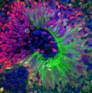
Every day, we call upon a unique capacity of our brain, visual mental imagery, which allows us to visualise images, objects or people 'in our heads'. Based on the recent case of a patient with a specific brain lesion, Paolo Bartolomeo's group (Inserm) in the PICNIC Lab at the Paris Brain Institute has identified a region that may be key in mental visualisation.
A patient was admitted to the emergency room after a stroke that had spread to the occipitotemporal area of the left hemisphere. Although his life was saved, the patient woke up with multiple deficits: hemianopia - the loss of vision on the right side - alexia - an inability to read - and an inability to name colours.
These multiple impairments and the presence of the lesion in the left temporal lobe prompted clinicians and researchers at the Paris Brain Institute to evaluate another brain function: visual mental imagery.
The brain networks of mental imagery
At present, the predominant model of the brain basis of mental imagery proposes that it engages the primary visual area at the back of our brain, which is also involved in processing what we actually see with our eyes. However, evidence from patient cases has been accumulating over the last twenty years that contradicts this dogma. In a recent meta-analysis, Paolo Bartolomeo's team suggested that mental imagery is instead encoded in the fronto-parietal networks of attention and working memory, as well as in a small region of the fusiform gyrus of the left temporal lobe. The case of this new patient with a left occipitotemporal lesion was therefore an opportunity for the researchers at the Paris Brain Institute to re-explore their hypothesis.
Intact visual mental imagery, despite the lesions
In order to test the patient's mental imagery, the doctors gave him a battery of tests. These consisted of several questions about the visual appearance of objects: What is redder between a strawberry and a cherry? Which city is the furthest on the right of a map of France between Bordeaux and Strasbourg? To answer correctly, the patient had to use his mental imagery and visualise in his head a strawberry, a cherry, or a map of France. "To our great surprise, our patient's visual mental imagery was well preserved," explains Paolo Bartolomeo (Inserm), the study's last author. "A new question then arose: why did he not have any difficulty, despite his lesion which should have affected important networks for exercising this function in our brain?”
Halfway between language and semantic networks
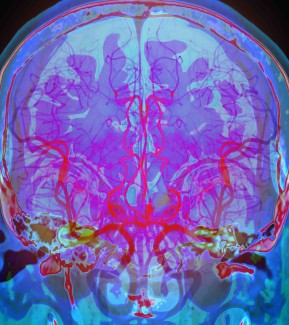
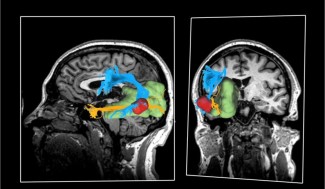
Thanks to MRI tractography, which makes it possible to visualise the tracts of neurons in the brain - the wiring, so to speak - the researchers were able to identify some key elements explaining why the patient's mental imagery was intact despite his lesion. They found that the mental imagery node, located in the fusiform gyrus in the left temporal lobe, had been spared by the lesion.
The team of scientists then showed that two connectivity tracts passed through this node: the arcuate fasciculus, associated with the language system, and the inferior longitudinal fasciculus, linked to the semantic system, i.e. our knowledge of the world, objects and concepts.
Because of his lesion, the patient no longer received direct visual information in his left hemisphere. The fusiform imagery node therefore no longer received this type of information but continued to be fed by the semantic network.
"These results support our hypothesis that visual mental imagery comes from a top-down activation from the language and semantic networks. This goes against the dominant model of mental imagery, according to which the primary visual areas are necessary for the implementation of this capacity" concludes Paolo Bartolomeo (Inserm).
Sources
https://pubmed.ncbi.nlm.nih.gov/35622159/
Hajhajate D, Kaufmann BC, Liu J, Siuda-Krzywicka K, Bartolomeo P. Brain Struct Funct. 2022 May 27