Immediate and secondary treatment for a stroke patient depends on the type of stroke, which is identified using an MRI scan. In 80% of cases, the stroke is caused by a blocked artery in the brain, referred to as a cerebral infarction. In 20% of cases, the stroke is caused by a rupture of a blood vessel in the brain, referred to as a cerebral hemorrhage.
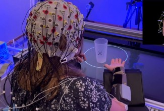
After a stroke and the emergence of focal damage, even if the underlying nerve tissue dies, the brain’s plasticity offers a chance of recovery. Thanks to neuroimaging techniques that use magnetic resonance imaging (MRI) in static states (observing the damage) or dynamic states (monitoring the activation of different regions of the brain during movement), we study the extent to which the different brain plasticity processes take place in each patient: the surrounding tissues may take over the lost function, the secondary areas involved in controlling a movement may become involved, or the contralateral areas (the healthy hemisphere not affected by the stroke) may take over, since we have two brain hemispheres. For example, when someone who has suffered a stroke shakes hands (if they are still able to), an area of their brain, different from that of a healthy individual, will be activated.
At Paris Brain Institute
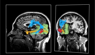
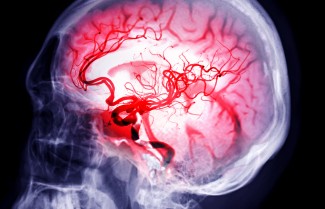
- Depending on the location of the residual brain damage from a stroke, the aftereffects will be different. This is why it is important to understand the structure of the brain vasculature. Nicolas Renier’s ‘Structural Dynamics of Networks’ team at Paris Brain Institute aims to map three dimensions of this vasculature in humans. The first step was the full mapping of the brain vasculature in an experimental model.
- From an evaluation of the aftereffects after the acute phase, up until ‘real-life’ rehabilitation, six caregivers, doctors and specialist researchers at Paris Brain Institute explain the challenges of complex post-stroke care and future pathways for improving patient recovery.