When a patient is admitted to intensive care due to a disorder of consciousness—such as a coma—establishing their neurological prognosis is a crucial yet challenging task. To reduce the uncertainty that precedes the medical decision, a group of clinicians and researchers from the Paris Brain Institute and the Pitié-Salpêtrière Hospital in Paris have assessed the efficacy of a multimodal approach that combines various clinical, electrophysiological, behavioural and neuroimaging indicators. The team's findings, published in Nature Medicine, show that this approach leads to better prognosis.
After a severe cranial trauma or cardiac arrest, some patients admitted to intensive care show little or no reaction to their environment—and are sometimes unable to communicate. This condition is called a disorder of consciousness (DoC), which includes comas, vegetative states and states of “minimal consciousness.”
This disorder sometimes persists for several days or weeks. In such cases, healthcare teams and relatives must obtain the most accurate information on the patient's cognitive recovery capacities. Usually, a neurological prognosis is established using several indicators—including standard measurements of brain anatomy (CT and MRI scans) and function (electroencephalogram).
“Despite having these data at our disposal, there remains a degree of uncertainty about the prognosis, which can significantly impact medical decision-making. These patients are often in a fragile state and prone to numerous complications, which raises questions about the appropriateness of the care they receive,” explains Benjamin Rohaut (AP-HP, Sorbonne University), neurologist, researcher and lead author of the study. “Moreover, doctors sometimes observe a discrepancy between the patient's behaviour and their brain activity: some patients in a vegetative state seem to understand what is being said to them but are unable to let their caregivers know.”
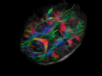
To improve the description of the state of consciousness of these patients, the “PICNIC team”, co-led by Lionel Naccache at the Paris Brain Institute, has been working for around fifteen years to define new brain measurements and clinical examination signs. Their approach has gradually evolved towards “multi-modality”, combining PET scans, multivariate EEG analysis, functional MRI, cognitive evoked potentials (electrical responses to sensory stimulation) and other tools.
Consciousness markers under scrutiny
To assess the clinical value of this approach, the team worked with the “Neurologically Oriented Intensive Care Unit” at the Pitié-Salpêtrière Hospital in Paris. Led by Benjamin Rohaut and Charlotte Calligaris (AP-HP), the clinicians and researchers followed and assessed 349 intensive-care patients between 2009 and 2021. At the end of each multimodal evaluation, they formulated a “good”, “uncertain”, or “unfavourable” prognostic opinion.
Their results indicate that patients with a “good prognosis” (22% of cases) showed a much more favourable evolution of their cognitive abilities than patients with a prognosis judged “uncertain” (45.5% of cases) or “unfavourable” (32.5% of cases); none of the patients assessed as “unfavourable” regained consciousness after one year. Above all, this prognostic performance was correlated with the number of modalities: the greater the number of indicators used, the greater the accuracy of the prognosis, and the greater the team's confidence in its assessments.
This long-term study shows for the first time the benefit of the multimodal approach, which is essential information for intensive care units worldwide. It also provides empirical validation of the recent recommendations of the European and American Neurology Academies.
However, the multimodal approach is not a magic wand. It provides the best possible information to caregivers and families in situations of uncertainty—an ethical advance in patient care—but does not guarantee bias-free decision-making.
Finally, there is the question of access to assessment tools, which are expensive and require specific expertise. “We are aware that multimodal assessment is not accessible to all the intensive care units that receive these patients”, continues Lionel Naccache (Sorbonne University, AP-HP). “We therefore propose to build a network of collaborations at the national and European levels. Thanks to telemedicine tools and automated EEG or brain imaging analysis, all intensive care units could have a first level of access to multimodal assessment. Should this prove insufficient, recourse to a regional expert centre would provide a more in-depth assessment. Finally, in the most complex situations, it would be possible to call on all available experts, wherever they may be. We aim to ensure that all patients with a disorder of consciousness can benefit from the highest standards of neurological prognosis.”
Funding
This study was funded by the James S. McDonnell Foundation, the Foundation for Medical Research (FRM), UNIM, the Lamonica Prize of the French Academy of Sciences, the European Partnership for Personalised Medicine (PerMed), and the Investissements d’avenir program.
Conflict of interest
Jacobo Sitt and Lionel Naccache are co-founders and shareholders of Neurometers, a company dedicated to the medical use of electroencephalogram (EEG) to quantify the brain signatures of consciousness and cognition.
Image
Crédit : Nicolas Decat.
Sources
Rohaut, B., Calligaris, C., … Sitt J.D., Naccache L. Multimodal assessment improves neuroprognosis performance in clinically unresponsive critical care patients with brain injury. Nature Medicine. (Mai 2024).
DOI : 10.1038/s41591-024-03019-1.

