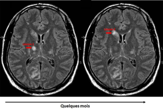
Multiple sclerosis diagnosis requires this combination of neurological symptoms with the presence of inflammatory plaques in an MRI scan, to see which have both spatial dissemination (brain, spinal cord, optic nerve) and temporal dissemination (inflammatory plaques of different ages or that appear over time).
Multiple sclerosis diagnosis
Diagnosing MS relies on an MRI scan detecting inflammatory plaques visible by a hypersignal in the brain and spinal cord, spread over time (recent and old lesions) and space (damage involving at least two regions of four possible locations in the central nervous system).
How multiple sclerosis evolves
The symptoms of the disease vary widely between patients. Similarly, the progression and time of onset of irreversible disability vary according to the ability of each affected person to ‘repair’ their brain damage.
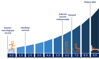
Whatever the type of multiple sclerosis, criteria are available to define the disease’s activity and monitor its evolution. Flare-ups, increases in the EDSS (Expanded Disability Status Scale) score (measuring disability severity) and the appearance of new damage that is visible on an MRI are recognized as markers of disease activity.
At Paris Brain Institute
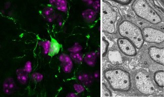
The team led by Profs. Catherine Lubetzki and Bruno Stankoff has found that the activation of microglia, the brain’s resident immune cells, in lesions is a reliable biomarker of the progression of patients’ disability. These results offer significant hope for adapting treatment for multiple sclerosis patients, evaluating new therapies, and reducing the severity of disability as much as possible.