Researchers from the Paris Brain Institute and Sainte-Justine University Hospital in Montreal have, for the first time, revealed the key stages of vascular development in the brain, from birth through adulthood. Using a 3D digital atlas called Lambada, they show that vascularization does not progress continuously, but instead unfolds in three distinct phases, closely linked to the maturation of neural circuits. These findings are published in Cell.
At birth, the brain is far from fully developed. Neural connections are formed at a rapid pace during the first weeks of life, and blood vessels—which supply oxygen and nutrients to nerve cells—follow a similar developmental trajectory. Until now, however, no one had described how blood vessels organize and adapt to neuronal needs on a day-to-day basis across the entire brain.
“The architecture of neural connections and the vascular network, once established, is largely preserved into adulthood,” explains Nicolas Renier (Inserm), head of the Plastic Lab at the Paris Brain Institute. “Many neurological diseases—whether neurodevelopmental disorders such as autism, cerebrovascular diseases, or even certain forms of epilepsy—are often associated with subtle disruptions in neurovascular construction. To understand how these diseases arise, it is essential to know what constitutes normal development. This reference did not exist.”
The mouse: a valuable model for studying vascular development
To address this gap, an international team led by Nicolas Renier at the Paris Brain Institute and Alexandre Dubrac at Sainte-Justine University Hospital set out to map the entire course of vascular development in the mouse brain, from birth to adulthood. The choice of this model is by no means insignificant.
“The mouse brain is highly immature at birth and resembles, in some respects, the fetal brain. The first two weeks of postnatal development in mice, therefore, model the last trimester of human brain development,” says Sophie Skriabine, co–first author of the study and researcher at University College London.
“The following weeks offer us a glimpse into the mechanisms of cerebral vascularization, from the final moments of fetal life through adolescence. In just three weeks, we observe the equivalent of about fifteen years of human development,” adds Elisa de Launoit, co–first author and postdoctoral researcher at the Francis Crick Institute.
Lambada: a goldmine for research
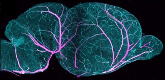
To build their atlas, the researchers used mouse brains made transparent using a chemical technique called iDISCO+. They then applied light-sheet microscopy—a method that scans tissues with a thin laser beam—to image blood vessels in every nook and cranny of the brain, down to the level of individual capillaries.
In total, the team performed more than 50 complete vascular reconstructions at nine different developmental stages, ranging from D3 (three days after birth) to D60 (the equivalent of a young adult).
Mapping the entire brain at high resolution over such a fine-grained time series was a real technological challenge that kept us busy for six years. In mice, the cerebral vascular network measures 40 meters at birth and reaches 300 meters by adolescence.
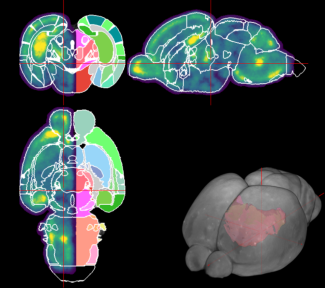
These three-dimensional images were then combined with spatial transcriptomics data, a technique that measures the activity of thousands of genes directly within tissues. The resulting atlas, Lambada (Lightsheet-Aligned Mouse Brain Annotated Developmental Atlas), is freely available online. It allows research teams worldwide to explore the developing brain in 3D, along with all associated molecular and anatomical data.
A symphony in three movements
The study’s findings challenge the traditional view of cerebral vascular development: rather than progressing uniformly, it unfolds in three clearly distinct stages.
Phase 1 – Uniform expansion (birth to D7).
During the first week of life, blood vessels grow homogeneously, in proportion to the increase in brain volume and its energy requirements, like scaffolding expanding alongside a building under construction. VEGF-A, a key vascular growth factor, plays a central role at this stage.
Phase 2 – Regional specialization (D7 to D21).
From day 7 onward, vessels begin to differentiate by brain region, supporting synapse formation and the emergence of brain functions. This period coincides with the mouse pup’s first sensory experiences: eyes open, whiskers grow and become sensitive, and hearing develops.
“In the very early stages of brain development, neurons fire in spontaneous bursts that carry no information about the outside world. Later on, we observe structured firing patterns linked to what the animal perceives in its environment,” notes Nicolas Renier. “Our data indicate that this transition from immature brain activity to sensory-driven activity coincides with the shift from phase 1 to phase 2 at the vascular level.”
Phase 3 – Stabilization (D21 to adulthood).
The network consolidates: unused vascular branches are eliminated, arteries reach maturity, and the final architecture of the adult brain takes shape. Astrocytes—glial cells that envelop blood vessels—appear to act as a “brake” to prevent excessive growth of the network, thereby contributing to the maturation of the blood–brain barrier.
A close dialogue between neurons and vessels
After describing these three phases, the researchers investigated their molecular environment. By cross-referencing vascular density maps with gene expression profiles for each brain region, they identified the molecules governing the interplay between neuronal activity and vascular growth at each developmental stage.
While VEGF-A dominates the first phase, other players take over in the second. Apelin and Wnt9a, two molecules associated with neuronal activity, appear to guide vascular densification where circuits need it most. Conversely, the protein Slit2 and angiotensinogen act as inhibitory signals, preventing excessive vascularization where it is unnecessary.
This integrated brake-and-accelerator system underlies the diversity of vascular organization in the brain and shapes blood vessel architecture as it matures and responds to the young mouse’s experiences.
Practical implications for biomedical research
Beyond these fundamental insights, Lambada is already establishing itself as a valuable resource for a wide range of research teams.
“Our data are being used by researchers studying hormone transport in the hypothalamic–pituitary complex during puberty, vascular recovery associated with hearing aid use in congenital deafness, and the effects of diet during development,” says Nicolas Renier.
In the longer term, the team aims to tackle a key question: how can early alterations in neurodevelopment weaken the brain’s vascular network and create conditions that predispose individuals to neurodegenerative diseases years later?
“A research project on the link between congenital hearing loss and the risk of developing dementia is already underway, in collaboration with Institut de l’audition in Paris. The research perspectives are immense,” concludes the researcher.
Sources
De Launoit, E., et al. The spatiotemporal dynamics of postnatal vascularization in the mouse brain. Brain. Avril 2026. DOI : 10.1016/j.cell.2026.03.013.
Funding
This study was funded by the French National Research Agency’s “Investissements d’avenir” program, the European Research Council, Alzheimer Research UK, and the Wellcome Leap Delta Tissue program.
Image
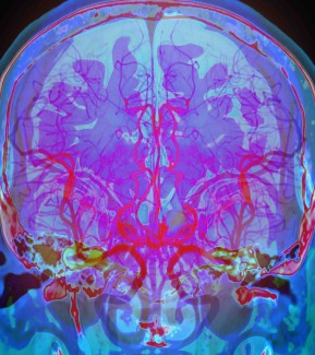
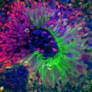
At birth, the brain has a rudimentary vascular network that becomes denser during the first weeks of postnatal life. Credit: Nicolas Renier.