
For decades, cell biology has built its approach around the study of genes and molecules, pushing the physical forces that sculpt our tissues into the background. Today, an international consortium, coordinated by Fanny Mochel at the Paris Brain Institute and Angeles Garcia-Cazorla at the Hospital Sant Joan de Déu in Barcelona, is calling for a shift of perspective whose implications could transform our understanding—and treatment—of brain diseases. The researchers’ reflections are published in the journal iScience.
Since the identification of DNA’s double helix in 1953, molecular biology has undergone a spectacular rise that has shaped our understanding of how cells function. Over the years, research has identified hundreds of genes involved in widespread neurological conditions such as Parkinson’s disease, as well as rarer disorders such as leukodystrophies.
Yet this success conceals a blind spot. Cells have gradually come to be regarded as mere biochemical systems—factories in which molecules assemble according to a program set by the genes. They are, however, also governed by the laws of physics, which exert forces on the tissues, fluids, or cell membranes, and underpin bioelectrical signaling.
This neglect of physics in brain research may partly explain the heterogeneous presentation of certain neurological diseases. “In inherited metabolic diseases, for example, we are faced with a puzzle: why do two patients carrying the same genetic mutation sometimes develop very different forms of the disease? Why do some show symptoms in childhood, and others only in adulthood? Genetic variants alone are not enough to account for this phenotypic variability”, explains Fanny Mochel (AP-HP, Sorbonne Université), co-leader of the MIND team at the Paris Brain Institute.
These are the questions that prompted Fanny Mochel, in collaboration with her counterpart, pediatric neurologist Angeles Garcia-Cazorla (Hospital Sant Joan de Déu) in Barcelona, to organize an international symposium of an unusual kind: Physics and Metabolism in Brain Functions. Now in its second edition, the event brings together physicists, chemists, philosophers, and neurobiologists to explore the mechanical properties of cells and tissues, and to assess the extent to which these may shed light on metabolic phenomena that genetics alone cannot describe.
When cells translate mechanical forces
A key concept shaping this new approach is mechanotransduction, the ability of cells to convert a mechanical signal—pressure, tension, or stretch—into a biochemical or electrical one. Examples abound. When the skin is injured, stretching of the epidermal cell membrane triggers a molecular cascade that recruits leukocytes to repair the tissue.
Receptors specialized for detecting mechanical forces, such as the ion channels PIEZO1 and PIEZO2, are found in many organs, including the stomach, lungs, bladder, and intestines. In the nervous system, they open when stretched or compressed, letting through a flow of calcium ions that alters the neuron’s electrical activity.
“In the same way, certain organelles are able to detect mechanical forces through the deformation of the cell’s membrane or of its cytoskeleton. They act, quite literally, as sensors”, Fanny Mochel notes. “And because these processes are essential to the exchange of signals and metabolites between cellular compartments, their disruption is associated with neurological diseases.”
Shaping the brain throughout life
Mechanical forces also sculpt brain development. They initiate the migration of neurons, the formation of axons, and the maturation of synapses, giving the cortex its characteristic folding into gyri and sulci. According to Eva Pillai, a cell biologist at EMBL, neural stem cells proliferate more in a soft environment; in a stiffer one, traction forces are transmitted through the cytoskeleton to the nucleus, opening nuclear pores and thereby altering gene expression.
“Tissue stiffness is a key concept for understanding the fate of the brain, both during development and during aging. Brain tissue is not made up only of neurons and glial cells: nearly 20% of its volume is occupied by the extracellular matrix—a meshwork of proteins (collagen, elastin), polysaccharides, and glycoproteins that surrounds the cells and gives them structure, elasticity, and viscosity. This matrix grows stiffer with age and with diseases”, adds Fanny Mochel.
Kevin Chalut, a biophysicist at Altos Labs in Cambridge, points out that oligodendrocyte progenitor cells—the stem cells that produce myelin—stop dividing in older subjects. But this is not an intrinsic decline: when aged cells are placed on a soft gel that mimics the highly pliant extracellular matrix of a young brain, they begin to proliferate again, and their gene expression profile rejuvenates.
In other words, it may be possible to act on aging itself by modifying the mechanical signals to which cells are exposed.
A revolution in tools
The promises of mechanomedicine, a medical approach that seeks to diagnose and treat diseases by acting on the mechanical properties of tissues, are all the more credible because they rest on innovative technologies.
“One example is magnetic resonance elastography, which makes it possible to measure tissue stiffness—a major source of information about how diseases manifest. The technique is still underused in brain imaging. And yet recent studies have shown that an increase in the stiffness of the medial temporal lobe predicts future cognitive decline in Alzheimer’s disease more accurately than classical measures of atrophy or amyloid burden”, explains Fanny Mochel.
Brillouin microscopy, for its part, now makes it possible to measure the stiffness of a tissue on a subcellular scale, without contact and without fluorescent labeling, by analyzing the interaction of light with the tissue’s internal acoustic vibrations. On the computational side, Marta Sales-Pardo (Rovira i Virgili University) has developed models capable of reconstructing the entire human metabolic network—over 13,000 chemical reactions—and projecting experimental data onto it to identify how a local perturbation, such as hyperglycemia, reshapes the system as a whole.
Adrien Hallou (University of Oxford) introduces an approach he calls spatial mechano-transcriptomics, which combines, for each cell in a mouse embryo, measurements of the mechanical forces acting on it with its gene expression profile. Finally, in Bordeaux, Laurent Cognet and his team are tracking, with the help of luminescent carbon nanotubes emitting in the near-infrared, the movement of individual molecules within the extracellular space of the living brain at a resolution finer than 50 nanometers.
Toward a mechanomedicine
If physical forces regulate cellular metabolism, they may also serve as levers for new therapies.
Several avenues are already being explored: hydrogels designed to restore an extracellular matrix stiffness favorable to neuronal regeneration; enzymes capable of stimulating synaptic plasticity; or artificial vesicles that target the transporters of the blood-brain barrier in order to promote the clearance of beta-amyloid peptides in Alzheimer’s disease.
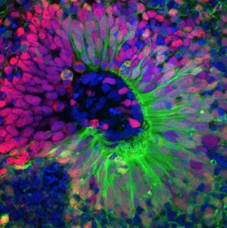
Stuart Hameroff, an anesthesiologist and researcher at the University of Arizona, proposes acting on microtubules—the cylindrical tubes that structure the interior of neurons—through transcranial ultrasound. In a pilot trial involving patients with chronic pain, the application of 8 MHz ultrasound improved mood, perhaps by interacting with the natural vibrations of microtubules. Sonogenetics, another emerging approach, consists of expressing mechanosensitive channels in selected neurons and then activating them with focused ultrasound, opening the way to non-invasive treatments for blindness or certain motor disorders.
Art as a thinking partner
Beyond the originality of these approaches, the very spirit of the consortium is to draw together disciplines, methods, and worldviews that tend to ignore one another in today’s hyper-specialized scientific culture.
“I am deeply convinced that, in order to solve complex problems in the field of brain diseases, we cannot confine ourselves to speaking among experts who share the same disciplinary culture, the same frames of reference”, argues Fanny Mochel. “It even seems necessary to me to work with non-scientists, who do not rely on the hypothetico-deductive method and who build practices and knowledge along other paths of thought, association, for instance. Artistic creation has a great deal to bring to science.”
To give shape to this ambition within the framework of the connections between physics, chemistry and neurobiology, Mochel and her colleagues draw on the concept of “night science”, coined by the French biologist and physician François Jacob in 1987. Night science is the exploration of hypotheses, ideas, and intuitions that are still incomplete—that have not yet taken the kind of form in which they can be put to the test through experiments.
This informal, sometimes secret way of working through scientific problems, outside the accepted methods, is nonetheless essential to scientific creativity. It will undoubtedly continue to inspire fresh approaches to the physical properties of living matter.
Night science wanders blindly. It hesitates, stumbles, falls back, sweats, wakes with a start. Doubting everything, it gropes for itself, questions itself, pulls itself together again and again. It is a kind of workshop of the possible, where what will one day become the material of science is being elaborated. Where hypotheses remain in the form of vague intimations, of misty sensations. Where phenomena are still nothing more than solitary events, with no link between them.
SOURCE
Mochel, F., Connections between physics and metabolism in brain functions. iScience, Février 2026. DOI : 10.1016/j.isci.2026.114643.
IMAGE
Microtubules in vitro. Crédit : Paul Appleton, Université de Dundee.
The team aims to understand the neuroinflammatory and metabolic mechanisms involved in multiple sclerosis and leukodystrophies, with a particular focus on the role of cellular metabolism and immune cells in lesion progression and central nervous...
Read more